Cell Structures & Organelles — Print · Grade 7 Science
Chapter: Cells, Transport & Cellular EnergyCell Structures & Organelles
Learning goals
- I can explain cell structures & organelles.
- I can use evidence, a model, and stated assumptions to support a biological explanation.
Before this unit
Use the supplied vocabulary, distinguish an observation from an explanation, and identify any units or model assumptions. Revisit a connected earlier unit if needed; new terms are explained here.
Use three flexible 40–45-minute sessions: core background and Parts 1–2 with their checks; Parts 3–4 and the worked application; then remaining practice, a labeled model, revision, and the exit check. Checks completed during reading can count toward the practice set. Read one short part at a time. Investigations and chapter review/quiz have separate sessions, and flexible weeks allow more time.
Your liver cells and your red blood cells carry the same DNA, and they do completely different jobs. What separates them is not their instructions but their equipment — what is actually built inside them. A cell is not a bag of soup. It is a crowded, organised system with a boundary, an interior, a store of information, and structures that each do particular work. The boundary is the cell membrane, and it is extraordinarily thin: about 8 nanometres, so roughly nine thousand of them stacked up would match the width of one human hair. Inside it is the cytoplasm, the jelly-like material that fills the cell and holds everything in place. Suspended in that cytoplasm are the organelles — the word means "little organs", and it is a fair description, because each one does a job the whole cell depends on, the way your organs do for you.
The largest organelle in most of your cells is the nucleus, and it is where almost all the cell's DNA is kept — the long molecule that carries the instructions for building proteins. The amount is hard to believe: unwound and laid end to end, the DNA from a single human cell would stretch about two metres, and it is packed into a nucleus roughly six micrometres across. Those instructions are not used in the nucleus itself. They are copied and carried out to the ribosomes, the small machines, about 20 nanometres wide, that assemble proteins by joining amino acids in the order the instructions specify. A busy cell can hold millions of ribosomes. Proteins matter because they do nearly everything: they form structures, speed up reactions, carry signals and move materials. The division of labour is worth remembering — the nucleus stores and protects the instructions, and the ribosomes build from them.
Doing all that work costs energy, and the organelles that release it are the mitochondria (one of them is a mitochondrion). During aerobic respiration they transfer energy stored in food molecules into a form the cell can spend, using oxygen and releasing carbon dioxide. Notice the verb: they transfer energy, they do not create it, and some of it always leaves as heat, which is one reason you are warm. A cell builds as many as it needs — a liver cell may hold one to two thousand, while a cell doing less work holds far fewer. Each mitochondrion is roughly a micrometre long, about the size of the bacteria from the last unit, which is a clue biologists have thought hard about. Two cautions before you meet a textbook diagram. Plant cells have mitochondria too and respire just as you do; having chloroplasts does not remove the need to release energy from food. And no single diagram fits every cell: a mature mammalian red blood cell has no nucleus and no mitochondria at all, having discarded both to make room for carrying oxygen.
Build your understanding
Where this fits
Last unit established that every living thing is made of cells and that new cells come from existing ones. That raises an obvious question: if a cell is the unit of life, what is inside one, and how do the parts work together? This unit opens the cell up. Keep the instrument limits from Chapter 1 in mind, because they decide what anyone could see and when.
Use three flexible 40–45-minute sessions: core background and Parts 1–2 with their checks; Parts 3–4 and the worked application; then remaining practice, a labeled model, revision, and the exit check. Checks completed during reading can count toward the practice set. Read one short part at a time. Investigations and chapter review/quiz have separate sessions, and flexible weeks allow more time.
Part 1 · The boundary that has to let things through
Start at the edge. The cell membrane is a double sheet of fat-like molecules called phospholipids, each with a water-loving head and two water-fearing tails. In water, those molecules line up automatically with the tails tucked inward and the heads facing out, which is why a membrane forms and reseals itself without anything having to assemble it. That double sheet is what makes the boundary possible, and it is only about 8 nanometres thick.
A sealed boundary would be useless. A cell has to take in glucose and oxygen and get rid of carbon dioxide and other products, so the membrane has to be selectively permeable — it lets some things through and holds others back. Small, uncharged molecules such as oxygen and carbon dioxide slip between the phospholipids on their own. Larger or charged particles, including glucose and sodium ions, cannot, and they cross through protein channels and pumps embedded in the sheet. The proteins are what make the selection specific.
Two things follow from that structure. First, what a cell can take in depends on which proteins it has built into its membrane, which is another way its equipment, not just its DNA, decides what it can do. Second, the membrane is not a decision-maker. Nothing in it chooses; molecules move according to physical and chemical rules, and the pattern we call regulation is the result. The plant cell wall you will meet in the next unit sits outside this membrane and does a different job entirely.
Pause and explain
Why would a completely sealed boundary be a poor model of a cell membrane?
Check your reasoning after answering
Because a sealed boundary would stop the exchanges the cell depends on. Oxygen and glucose could not come in and carbon dioxide could not leave, so respiration would stop. A real membrane is selectively permeable: small uncharged molecules cross it directly, and larger or charged ones pass through specific proteins.
Part 2 · Where the instructions are kept, and where proteins are built
The nucleus is a compartment with its own double membrane, pierced by pores, and it holds the cell's DNA. Keeping the instructions separated from the rest of the cell has a real advantage: the copy that leaves can be controlled, so the cell can make a lot of one protein and none of another without altering the original. The nucleus does not build anything. It stores, protects and issues.
Ribosomes do the building. A ribosome reads a copied instruction and joins amino acids in the specified order, producing a protein that then folds into a shape determined by that order. Some ribosomes float free in the cytoplasm; others sit on a folded membrane system called the endoplasmic reticulum, and those tend to make proteins destined to leave the cell. A cell that secretes a great deal, such as a cell in your pancreas, is crowded with them.
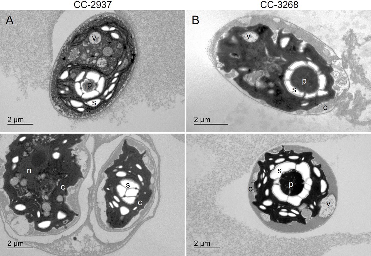
This is also where the division between the two kinds of cell matters. Eukaryotes — animals, plants, fungi, and the single-celled Paramecium from the last unit — keep their DNA inside a nucleus. Prokaryotes, the bacteria, do not: their DNA sits directly in the cytoplasm. But prokaryotes still have DNA, still have ribosomes, and still build proteins. A diagram showing a tidy nucleus is a model of one kind of cell, not an inventory of life.
Pause and explain
Does the nucleus itself assemble the cell's proteins?
Check your reasoning after answering
No. The nucleus stores the DNA instructions; ribosomes assemble proteins by joining amino acids in the order a copied instruction specifies. Separating the two lets the cell control how much of each protein it makes. Bacteria have no nucleus at all and still build proteins, because they still have DNA and ribosomes.
Part 3 · Releasing energy from food, and why 'making energy' is wrong
A mitochondrion is roughly a micrometre long and has two membranes, the inner one folded into deep ridges. Those folds matter: they pack a large working surface into a small volume, and most of the energy transfer happens on them. During aerobic respiration the mitochondrion uses oxygen to break down glucose, releasing carbon dioxide and water and transferring the energy into a molecule the cell can spend wherever it is needed.
The phrase "mitochondria make energy" is the most common error in this unit, and it is worth being precise about why it is wrong. Energy is not created; it is moved from one store to another. The chemical energy was already in the glucose, put there by a plant. The mitochondrion transfers part of it into a usable form and the rest escapes as thermal energy to the surroundings. That escaping fraction is not waste in any useless sense — it is why a mammal is warm to the touch.
Cells build the number of mitochondria their work demands. A liver cell or a heart muscle cell, both working constantly, may hold one to two thousand. A mature mammalian red blood cell holds none: it discards its nucleus and mitochondria as it matures so that more of its volume can carry oxygen, and it survives on a simpler process that needs neither. Plant cells, which make their own glucose, still respire in mitochondria to use it.
Pause and explain
Why is "mitochondria create energy" misleading?
Check your reasoning after answering
Because energy cannot be created. The chemical energy is already stored in glucose; the mitochondrion transfers part of it into a form the cell can spend and loses the rest as heat. Saying a structure creates energy hides where the energy came from, which is food, and ultimately a plant that captured sunlight.
Part 4 · Explaining a system instead of labelling a diagram
Naming organelles is the easy half. An explanation connects them: the membrane admits glucose and oxygen; the nucleus issues an instruction; a ribosome builds the protein that instruction specifies; a mitochondrion supplies the usable energy that the building costs; and the proteins produced go on to form structures and run reactions, including the channels in the membrane where the chain started. That loop is the cell as a system.
The test of whether you have an explanation rather than a list is a damage question. If a cell's mitochondria stop working, protein building slows, membrane pumps that need energy fail, and internal conditions drift — not because the mitochondria did those jobs, but because the work they paid for cannot be paid for any more. If instead the membrane is torn, the contents leak and conditions cannot be maintained, even though every organelle inside may be perfectly intact.
That second case is why a cell is not a collection of independent parts. An organelle is only as useful as the system it sits in. When you read a claim about a cell, ask which structure is doing what, what it needs from the others, and what evidence shows the connection — rather than accepting a label as an explanation.
Pause and explain
Why is labelling organelles not yet an explanation of how a cell works?
Check your reasoning after answering
Because labels name parts without saying what depends on what. An explanation states each structure's role and the connections between them: the membrane supplies materials, the nucleus issues instructions, ribosomes build proteins, mitochondria supply the energy that building costs, and the proteins made then maintain the membrane.
Apply the whole unit · Use structure and function as evidence
Illustrative cell record, not a measured experiment: a cell's membrane has been damaged and substances are leaking out of it. Under the microscope the ribosomes are still clearly visible and look normal. A student's draft concludes that because the ribosomes are intact, the cell is still functioning normally.
Your task
Explain what the membrane does, evaluate the draft's reasoning, and name one further observation that would settle the question.
What your response should show
- Explain the relevant biological process accurately.
- Use the supplied evidence and show calculation steps with units where needed.
- Connect a labeled model or mechanism with the claim.
- State an assumption, evidence limitation, or useful next observation.
Response guidance — after your attempt
State the membrane's role: it is the selective boundary that admits glucose and oxygen, removes products, and maintains internal conditions different from the surroundings.
Say what intact ribosomes do and do not show. They show that one structure survived; they do not show that it has the amino acids, instructions or energy it needs to work.
Identify the reasoning error: the draft treats one intact part as evidence about the whole system, when the damaged part is the one every other part depends on.
Name a further observation: whether the cell is still making protein, still maintaining a different internal concentration from its surroundings, or still consuming oxygen — each tests function rather than appearance.
The membrane is the selective boundary. It lets oxygen and glucose in, lets carbon dioxide out, and keeps the inside of the cell chemically different from the outside; a cell that cannot do this cannot maintain the conditions its reactions need. The draft's reasoning does not hold. Visible, normal-looking ribosomes establish that one organelle is structurally intact at the moment of observation, not that it is still building proteins, and certainly not that the cell as a whole is functioning. With the boundary breached, materials leak out, concentrations equalise with the surroundings, and the energy supply that protein building depends on is interrupted — so an intact ribosome may simply be an intact ribosome in a dying cell. A single further observation would settle it: test whether the cell is still producing protein, still consuming oxygen, or still holding an internal concentration different from the fluid around it. Any of those measures function directly, whereas appearance under a microscope measures only structure, and only at one instant.
Scientific references for this unit
Parent / teacher teaching notes
Ask for a because-statement and a labeled relationship, not just a memorized word. Work through one calculation step at a time, permit oral or diagram responses, and use flexible sessions for difficult reading. Compare the learner’s actual evidence with the supplied model; do not reward copying or agreement with a personal preference. No private family, genetic, or medical records are required.
The key idea
A cell is an interacting system with boundaries, internal material, genetic information, and structures that perform tasks. The membrane surrounds the cell and helps regulate exchanges. Cytoplasm contains material and structures inside the membrane. An organelle is a specialized structure within a cell; many familiar organelles occur in eukaryotes, while prokaryotes lack a membrane-enclosed nucleus.
Worked examples
Example 1 · Read equipment, not instructions
- Separate instructions from equipment: identical DNA means the same set of possible proteins, not the same structures actually built.
- Match each cell's equipment to its job. Heart muscle contracts without pause and needs a constant supply of usable energy, so it builds mitochondria in large numbers.
- A red blood cell's job is to carry oxygen. As it matures it discards its nucleus and mitochondria, freeing volume for haemoglobin and avoiding consuming the oxygen it is meant to deliver.
Same instructions, different equipment — that is the whole reason one body can contain cells that behave nothing alike.
Example 2 · Follow a chain of dependence
- Identify what mitochondria supply: not proteins, but the usable energy that other processes spend.
- Find the processes that spend it. Building a protein means joining amino acids, which costs energy, and so does pumping materials across the membrane to collect those amino acids.
- Trace the consequence rather than assuming an organelle stops working on its own: ribosomes are intact but under-supplied, so production slows and then stops.
Ask what each structure supplies to the others, not just what it is called.
Avoid these mistakes
- Mistake: mitochondria make energy. They transfer energy already stored in glucose into a usable form and lose some as heat; the energy came from food, and before that from a plant capturing sunlight.
- Mistake: the nucleus builds proteins. It stores and issues the DNA instructions, while ribosomes do the building — which is why bacteria, with no nucleus, still make proteins.
- Mistake: every cell contains every organelle in the diagram. A mature mammalian red blood cell has neither nucleus nor mitochondria, and a bacterium has no membrane-enclosed nucleus.
- Mistake: a labelled diagram is an explanation. Naming parts is not saying what depends on what; an explanation connects membrane, instructions, ribosomes and energy supply into one working system.
Check your understanding
Why would a completely sealed boundary be a poor cell model?
Discuss the answer
It would prevent needed exchanges such as uptake of materials and removal of wastes.
Does a nucleus itself assemble all proteins?
Discuss the answer
No. Ribosomes build proteins using cellular instructions; the nucleus stores much genetic information in a typical eukaryotic cell.
Words to know
- organelle
- A structure inside a cell that does a particular job; the word means "little organ".
- cytoplasm
- The jelly-like material filling the cell inside its membrane, holding the organelles in place.
- nucleus
- The compartment holding almost all of a eukaryotic cell's DNA; it stores and issues instructions rather than building anything.
- ribosome
- A machine about 20 nanometres across that builds a protein by joining amino acids in the order an instruction specifies.
- mitochondrion
- The organelle, about a micrometre long, that transfers energy from food into a usable form during aerobic respiration.
Extend your scientific explanation
Compare a second supplied example or the linked educational reference. Identify a connection, a model limit, and an additional observation that would test the explanation.
In the real world
Use a clear question, reliable observations, and a biological mechanism to evaluate a claim. State what remains uncertain before applying a classroom model to a different setting.
Exit check
Why is labeling organelles not a complete explanation of cell function?
Check your explanation after answering
An explanation also connects each structure with its role and interactions in the functioning system.
Explain your reasoning without notes. If a key idea is missing, revisit the lesson and try again.
Problem 1
Answer and explanation
It would prevent needed exchanges such as uptake of materials and removal of wastes.
Problem 2
Answer and explanation
No. Ribosomes build proteins using cellular instructions; the nucleus stores much genetic information in a typical eukaryotic cell.
Problem 3
Answer and explanation
They help transfer energy from food into usable forms; energy is not created.
Problem 4
Answer and explanation
An explanation also connects each structure with its role and interactions in the functioning system.
Problem 5
Answer and explanation
The membrane helps maintain regulated exchanges and internal conditions. Leakage suggests disruption even though ribosomes remain visible. Presence of one structure does not prove normal system function. Measurements of exchange, internal conditions, and cell activity would help assess the extent of damage.
Locate the exchange boundary.
Connect uncontrolled leakage with disrupted internal conditions.
Separate visible presence from measured function.
Response criteria
- Explain the relevant biological process accurately.
- Use the supplied evidence and show calculation steps with units where needed.
- Connect a labeled model or mechanism with the claim.
- State an assumption, evidence limitation, or useful next observation.
Problem 6
Answer and explanation
Yes. Ribosomes can use cellular instructions without an enclosed nucleus; the arrangement differs from the typical eukaryotic-cell model.
Response criteria
- Explain the relevant biological process accurately.
- Use the supplied evidence and show calculation steps with units where needed.
- Connect a labeled model or mechanism with the claim.
- State an assumption, evidence limitation, or useful next observation.
Problem 7
- Cell wall
- Ribosome
- Scale bar
- Whole organ system
Answer and explanation
Ribosome
Ribosomes assemble proteins using cellular instructions.
Problem 8
Answer and explanation
Building, transporting, repairing, and maintaining cellular conditions require energy transfers. Photosynthesis stores energy in food; respiration releases energy into usable forms.